KPV Peptide: Mechanism, Anti-Inflammatory Research & Scientific Overview
A comprehensive scientific review of KPV peptide, the C-terminal tripeptide fragment of alpha-MSH, covering its molecular structure, NF-kB inhibition mechanisms, and current research findings in inflammation and gut health studies.
KPV Peptide: Mechanism, Anti-Inflammatory Research & Scientific Overview
Key Points
- KPV is a C-terminal tripeptide fragment (Lys-Pro-Val) derived from alpha-melanocyte-stimulating hormone (alpha-MSH)
- Molecular formula: C16H30N4O4 with a molecular weight of 342.43 g/mol
- Research indicates involvement in NF-kB inhibition and anti-inflammatory pathways
- Studies conducted primarily in animal models and in vitro cell culture systems
- Not approved by FDA for human therapeutic use; remains an emerging research compound
Table of Contents
- Introduction
- Molecular Structure
- Mechanism of Action
- Research Overview
- Stability & Handling
- Current Research Limitations
- Conclusion
- References
Introduction
KPV is a naturally occurring tripeptide consisting of three amino acids: lysine (K), proline (P), and valine (V). This peptide represents the C-terminal fragment (amino acids 11-13) of alpha-melanocyte-stimulating hormone (alpha-MSH), a 13-amino acid neuropeptide produced by cells in the hypothalamus, pituitary gland, and various peripheral tissues.
Alpha-MSH has long been recognized for its role in pigmentation regulation through melanocortin receptor activation. However, research beginning in the 1980s and 1990s identified additional properties of this hormone, including immunomodulatory and anti-inflammatory effects. Subsequent investigations revealed that the C-terminal KPV sequence retained certain biological activities independent of melanocortin receptor binding, suggesting alternative mechanisms of action.
The isolation and characterization of KPV as a distinct bioactive fragment emerged from efforts to understand which portions of the alpha-MSH molecule contributed to its various observed effects. Unlike the full-length hormone, KPV does not activate classical melanocortin receptors (MC1R-MC5R) at physiologically relevant concentrations, indicating that its documented activities operate through different molecular pathways.
This article provides an objective examination of current KPV research findings, focusing on documented mechanisms and experimental observations in preclinical models. It is important to note that KPV remains an early-stage research compound with limited human clinical data, and the findings discussed represent emerging science requiring further validation.
Molecular Structure
Chemical Properties
| Property | Value |
|---|---|
| Molecular Formula | C16H30N4O4 |
| Molecular Weight | 342.43 g/mol |
| Sequence | Lys-Pro-Val (H-Lys-Pro-Val-OH) |
| Amino Acid Count | 3 |
| CAS Number | 67727-97-3 |
| Isoelectric Point | ~9.7 |
Structural Characteristics
KPV is characterized by its simple tripeptide structure, representing one of the smallest bioactive peptide fragments studied in anti-inflammatory research. The sequence begins with lysine, a basic amino acid with a positively charged epsilon-amino group at physiological pH, followed by proline, which introduces a rigid kink in the peptide backbone, and terminates with valine, a hydrophobic branched-chain amino acid.
The presence of proline in the central position is structurally significant. Proline is the only amino acid where the side chain is bonded to the backbone nitrogen, creating a cyclic structure that restricts conformational flexibility. This characteristic may contribute to the peptide's stability and its specific interactions with cellular targets.
Relationship to Alpha-MSH
Alpha-MSH (also designated alpha-melanotropin) has the complete sequence: Ac-Ser-Tyr-Ser-Met-Glu-His-Phe-Arg-Trp-Gly-Lys-Pro-Val-NH2. The KPV tripeptide corresponds to positions 11, 12, and 13 of this sequence. While the N-terminal and central portions of alpha-MSH are primarily responsible for melanocortin receptor activation and pigmentary effects, the C-terminal KPV region has been associated with receptor-independent immunomodulatory activities.
Research has demonstrated that KPV can be generated endogenously through proteolytic processing of alpha-MSH by various peptidases present in inflammatory environments. This suggests a potential physiological role for this fragment in modulating inflammatory responses, though the extent and significance of endogenous KPV production remains under investigation.
Mechanism of Action
Research suggests KPV interacts with cellular signaling pathways through mechanisms distinct from classical melanocortin receptor activation. The following pathways have been documented in preclinical studies:
NF-kB Inhibition
The primary mechanism attributed to KPV involves inhibition of nuclear factor kappa-B (NF-kB), a transcription factor central to inflammatory gene expression. NF-kB regulates the transcription of numerous pro-inflammatory mediators, including cytokines (IL-1, IL-6, TNF-alpha), chemokines, and adhesion molecules.
Studies by Brzoska et al. and subsequent investigators have demonstrated that KPV can:
- Inhibit IkB kinase (IKK) activity, preventing phosphorylation and degradation of IkB-alpha
- Reduce nuclear translocation of NF-kB p65/p50 subunits
- Decrease DNA binding activity of NF-kB to target gene promoters
- Downregulate expression of NF-kB-dependent inflammatory genes
Research in various cell types, including intestinal epithelial cells, macrophages, and keratinocytes, has consistently demonstrated NF-kB pathway modulation following KPV exposure, though the precise molecular target through which KPV initiates this inhibition remains under investigation.
Intracellular Peptide Transport
A distinctive feature of KPV pharmacology is its proposed mechanism of cellular entry. Research by Kannengiesser et al. (2007) demonstrated that KPV utilizes the oligopeptide transporter PepT1 (SLC15A1) to enter intestinal epithelial cells. PepT1 is a proton-coupled transporter that mediates uptake of di- and tripeptides across the intestinal brush border membrane.
This transport mechanism has several implications:
- Oral bioavailability potential: Unlike most peptides that require injection, PepT1-mediated uptake suggests possible oral absorption of KPV
- Tissue specificity: PepT1 expression is highest in the small intestine, potentially concentrating KPV effects in gastrointestinal tissue
- Intracellular localization: Entry via PepT1 delivers KPV directly to the cytoplasm where it can interact with signaling components
Studies have confirmed that inhibition of PepT1 blocks KPV uptake and abolishes its anti-inflammatory effects in intestinal epithelial cell models, supporting the importance of this transport pathway.
Inflammasome Modulation
Emerging research suggests KPV may influence inflammasome activity, particularly the NLRP3 inflammasome complex. Inflammasomes are intracellular multiprotein complexes that activate caspase-1 and promote maturation and secretion of IL-1beta and IL-18.
Preliminary studies have indicated that KPV exposure may:
- Reduce NLRP3 inflammasome assembly
- Decrease caspase-1 activation
- Lower IL-1beta secretion in stimulated macrophages
However, this area of KPV research remains in early stages, and the mechanisms linking NF-kB inhibition to inflammasome effects require further clarification.
Melanocortin Receptor Independence
It is important to emphasize that KPV's anti-inflammatory activities appear largely independent of melanocortin receptor activation. Studies have demonstrated:
- KPV does not bind MC1R with significant affinity at concentrations showing anti-inflammatory effects
- Anti-inflammatory effects persist in MC1R-deficient models
- The activity profile differs from classical melanocortin receptor agonists
This receptor independence distinguishes KPV from other alpha-MSH-derived peptides and suggests a distinct pharmacological profile.
Research Overview
Inflammatory Bowel Disease Models
The most extensively studied application of KPV involves inflammatory bowel disease (IBD) models. Research in this area has been driven by the combination of KPV's anti-inflammatory mechanism and its uptake via intestinal PepT1 transporters.
Colitis Model Studies
- Laroui et al. (2010) demonstrated reduced inflammation in DSS-induced colitis models following KPV administration
- Studies showed decreased histological damage scores and reduced inflammatory cell infiltration
- Cytokine analysis revealed lower levels of TNF-alpha, IL-6, and IL-1beta in colonic tissue
- KPV-loaded nanoparticles showed enhanced efficacy compared to free peptide in some studies
Proposed Mechanisms in Gut Inflammation
Research suggests KPV may affect intestinal inflammation through:
- Direct uptake by epithelial cells via PepT1 and subsequent NF-kB inhibition
- Preservation of epithelial barrier integrity
- Modulation of inflammatory cell activation in lamina propria
- Potential effects on gut microbiome composition (emerging area of investigation)
Skin Inflammation Research
Given alpha-MSH's established role in skin biology, researchers have examined KPV in dermatological inflammation models.
In Vitro Studies
- Keratinocyte cultures showed reduced IL-8 and TNF-alpha production following KPV treatment
- UV-irradiated skin cell models demonstrated decreased inflammatory marker expression
- Studies in melanocyte cultures indicated potential cytoprotective effects
Animal Model Research
- Contact dermatitis models showed modified inflammatory responses with topical KPV application
- Wound healing studies indicated effects on inflammatory phase duration
- Limited research in psoriasis-like models has shown preliminary effects on keratinocyte proliferation markers
Antimicrobial Properties
An intriguing area of KPV research involves its documented antimicrobial activities, which are independent of its anti-inflammatory mechanisms.
Observed Antimicrobial Effects
- Direct antimicrobial activity against Staphylococcus aureus and Candida albicans demonstrated in vitro
- Mechanisms may involve membrane disruption and/or interference with microbial signaling
- The lysine residue's positive charge likely contributes to membrane interactions
Research Implications
The combination of anti-inflammatory and antimicrobial properties has generated interest in KPV's potential for addressing infections with inflammatory components, though this remains a preclinical research area.
Neuroinflammation Studies
Limited but growing research has examined KPV in neuroinflammatory contexts:
- Microglial activation studies showed reduced pro-inflammatory cytokine production
- Blood-brain barrier permeability research has explored potential CNS access
- Neuroprotection studies in oxidative stress models have shown preliminary results
This area requires substantially more investigation before drawing conclusions about neurological applications.
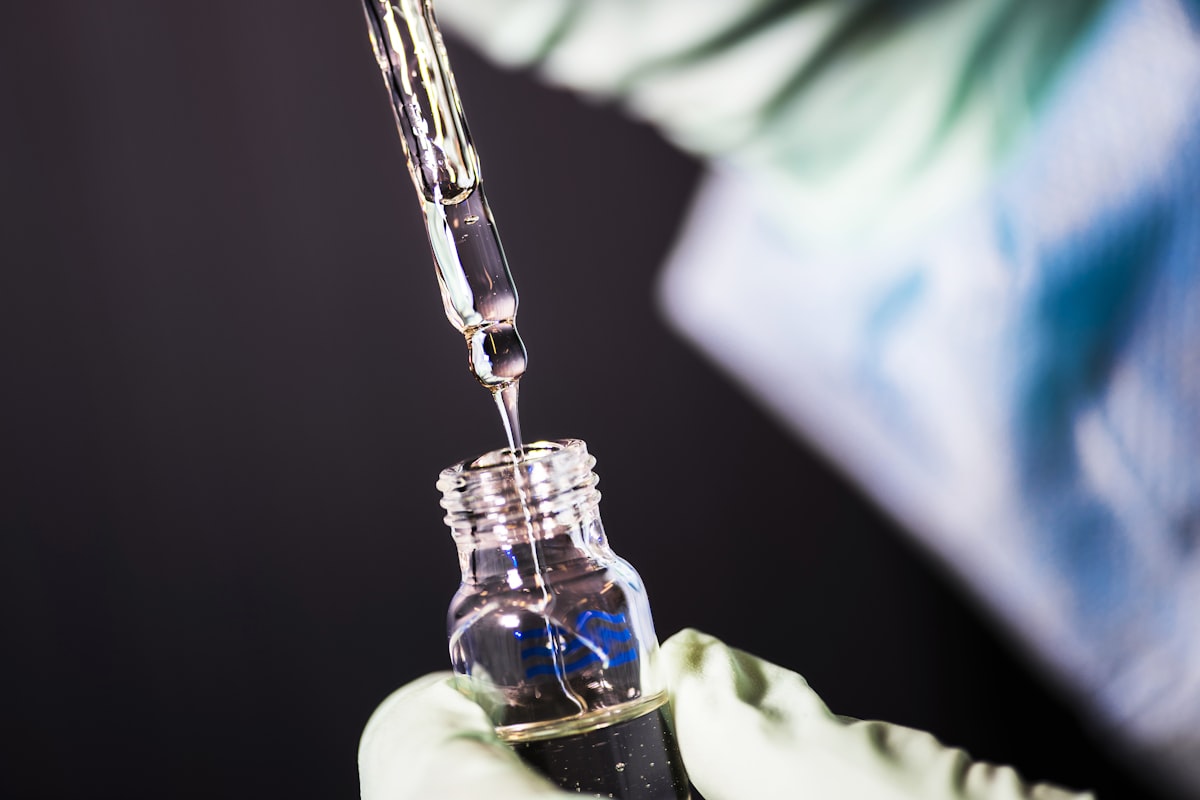
Stability & Handling
Storage Requirements
| Condition | Recommendation |
|---|---|
| Lyophilized Form | -20C, protected from light, stable 2+ years |
| Reconstituted (Bacteriostatic Water) | 2-8C, use within 4 weeks |
| Reconstituted (Sterile Water) | 2-8C, use within 1-2 weeks |
| Working Solutions | Prepare fresh, avoid repeated freeze-thaw cycles |
Reconstitution Guidelines
For research applications:
- Allow lyophilized peptide to reach room temperature before opening
- Add reconstitution solvent slowly along vial wall to prevent foaming
- Swirl gently until dissolved; do not vortex aggressively
- Filter sterilize if required for cell culture applications
- Aliquot to minimize freeze-thaw exposure
- Document concentration and storage conditions
Stability Considerations
KPV demonstrates favorable stability characteristics for a small peptide:
- pH Stability: Maintains integrity across pH range 4-8
- Thermal Stability: Relatively stable at room temperature for short periods
- Protease Resistance: The proline residue provides some protection against aminopeptidases
- Oxidation Sensitivity: Low compared to methionine- or cysteine-containing peptides
The small size and lack of oxidation-sensitive residues contribute to KPV's practical advantages in research settings. However, as with all peptides, proper storage and handling protocols should be followed to ensure consistent experimental results.
Current Research Limitations
Study Quality Considerations
Critical evaluation of KPV research reveals several important limitations:
-
Predominantly Preclinical Data: The vast majority of studies have been conducted in cell culture systems and animal models, with minimal human clinical data available
-
Limited Independent Replication: While multiple research groups have studied KPV, comprehensive independent replication of key findings remains limited
-
Dosing Variability: Studies have employed widely varying KPV concentrations, complicating cross-study comparisons and dose-response characterization
-
Mechanistic Gaps: The precise molecular target(s) through which KPV initiates NF-kB inhibition have not been definitively identified
-
Pharmacokinetic Data: Detailed absorption, distribution, metabolism, and excretion (ADME) profiles in relevant species are incomplete
Human Research Status
As of 2026, KPV remains in early research stages:
- No completed clinical trials in peer-reviewed literature
- No FDA investigational new drug (IND) applications publicly documented
- No regulatory approval for any therapeutic indication
- Classified strictly as a research compound
Species Translation Concerns
Several factors complicate translation of animal findings to human biology:
- PepT1 expression patterns may differ between species
- Inflammatory pathway regulation varies across species
- Endogenous alpha-MSH processing may differ
- Gut physiology and microbiome composition are species-specific
Areas Requiring Further Investigation
The following research gaps should be addressed before KPV can advance toward clinical applications:
- Identification of direct molecular targets
- Comprehensive pharmacokinetic profiling in multiple species
- Long-term safety assessment studies
- Dose-response characterization in standardized models
- Investigation of potential drug interactions
- Assessment of immunogenicity and tolerance
- Human tissue and ex vivo validation studies
Research Peptides
Lab-verified purity with full COA documentation. Wholesale pricing for research institutions.
Conclusion
KPV represents an emerging research compound with documented anti-inflammatory properties in preclinical models. Its proposed mechanism involving NF-kB pathway inhibition, combined with PepT1-mediated intestinal uptake, has generated particular interest in gastrointestinal inflammation research. Additional studies have explored its effects in skin inflammation models and documented antimicrobial properties.
As a fragment of alpha-MSH, KPV provides insights into how larger bioactive peptides can be processed into smaller fragments with distinct pharmacological profiles. Its receptor-independent mechanism differentiates it from classical melanocortin system modulators and suggests alternative therapeutic strategies may be possible.
However, the current body of evidence consists primarily of in vitro and animal model studies. Critical gaps remain in understanding KPV's precise molecular targets, pharmacokinetic behavior, and translational potential to human disease. The absence of clinical trial data and regulatory approval underscores that KPV remains a research tool for investigating inflammatory mechanisms rather than an established therapeutic agent.
Researchers utilizing KPV should approach the literature critically, recognizing both the promising preliminary findings and the substantial additional work required to validate these observations in human contexts. As with all emerging research compounds, conclusions should be tempered by the early stage of scientific investigation.
Browse Our Peptide Catalog
View our full range of research peptides with COA documentation and purity specs.
References
-
Brzoska T, Luger TA, Maaser C, et al. Alpha-melanocyte-stimulating hormone and related tripeptides: biochemistry, antiinflammatory and protective effects in vitro and in vivo, and future perspectives for the treatment of immune-mediated inflammatory diseases. Endocr Rev. 2008;29(5):581-602. doi:10.1210/er.2007-0027
-
Kannengiesser K, Maaser C, Heidemann J, et al. Melanocortin-derived tripeptide KPV has anti-inflammatory potential in murine models of inflammatory bowel disease. Inflamm Bowel Dis. 2008;14(3):324-331. doi:10.1002/ibd.20334
-
Laroui H, Dalmasso G, Nguyen HTT, et al. Drug-loaded nanoparticles targeted to the colon with polysaccharide hydrogel reduce colitis in a mouse model. Gastroenterology. 2010;138(3):843-853.e2. doi:10.1053/j.gastro.2009.11.003
-
Catania A, Gatti S, Colombo G, Lipton JM. Targeting melanocortin receptors as a novel strategy to control inflammation. Pharmacol Rev. 2004;56(1):1-29. doi:10.1124/pr.56.1.1
-
Dalmasso G, Charrier-Hisamuddin L, Nguyen HTT, et al. PepT1-mediated tripeptide KPV uptake reduces intestinal inflammation. Gastroenterology. 2008;134(1):166-178. doi:10.1053/j.gastro.2007.10.026
-
Luger TA, Brzoska T. Alpha-MSH related peptides: a new class of anti-inflammatory and immunomodulating drugs. Ann Rheum Dis. 2007;66(Suppl 3):iii52-iii55. doi:10.1136/ard.2007.079780
-
Maaser C, Kannengiesser K, Kucharzik T. Role of the melanocortin system in inflammation. Ann N Y Acad Sci. 2006;1072:123-134. doi:10.1196/annals.1326.016
-
Getting SJ, Christian HC, Lam CW, et al. Redundancy of a functional melanocortin 1 receptor in the anti-inflammatory actions of melanocortin peptides: studies in the recessive yellow (e/e) mouse suggest an important role for melanocortin 3 receptor. J Immunol. 2003;170(6):3323-3330. doi:10.4049/jimmunol.170.6.3323
-
Cutuli M, Cristiani S, Lipton JM, Bhardwaj N. Antimicrobial effects of alpha-MSH peptides. J Leukoc Biol. 2000;67(2):233-239. doi:10.1002/jlb.67.2.233
-
Capsoni F, Carrabba M, Braga M, et al. Melanocortin peptides inhibit prostaglandin E2 release by human osteoarthritis synovial tissue. Ann Rheum Dis. 2002;61(2):185-188. doi:10.1136/ard.61.2.185
-
Bohm M, Luger TA. Melanocortins in fibroblast biology--current update and future perspective for dermatology. Exp Dermatol. 2004;13(Suppl 4):16-21. doi:10.1111/j.1600-0625.2004.00257.x
-
Yoon SW, Chun JS, Sung MH, Kim JY, Poo H. Alpha-MSH inhibits TNF-alpha-induced matrix metalloproteinase-13 expression by modulating p38 kinase and nuclear factor kappaB signaling in human chondrosarcoma HTB-94 cells. Osteoarthritis Cartilage. 2008;16(1):115-124. doi:10.1016/j.joca.2007.05.026
-
Mykicki N, Herrmann AM, Schwab N, et al. Melanocortin-1 receptor activation is neuroprotective in mouse models of neuroinflammatory disease. Sci Transl Med. 2016;8(362):362ra146. doi:10.1126/scitranslmed.aaf8732
-
Rajora N, Boccoli G, Burns D, et al. Alpha-MSH modulates local and circulating tumor necrosis factor-alpha in experimental brain inflammation. J Neurosci. 1997;17(6):2181-2186. doi:10.1523/JNEUROSCI.17-06-02181.1997
Reviewed by: Dr. Research Reviewer, PhD